Imådje:Brain - Lobes.png
I n’ a nén di pus grande finté.
Brain_-_Lobes.png (701 × 487 picsels, groxheur do fitchî: 360 Ko, del sôre "MIME": image/png)
Ci fitchî ci provént d’ Wikimedia Commons ; si pout i esse eployî divins des ôtes pordjets Wiki. Li discrijhaedje di si pådje di discrijhaedje låvå est håynêye cial ådzo.
| Discrijhaedje |
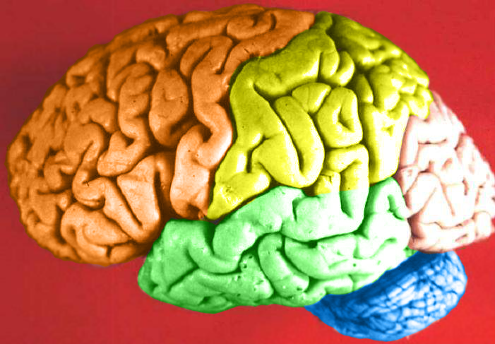
Human brain lateral view - Lobes
|
| Date | (UTC) |
| Sourdant | Human_brain_lateral_view_description.JPG |
| Oteur | Dep't. of Cellular Biology & Anatomy, Louisiana State University Health Sciences Center Shreveport |
| Otorijhaedje (Reployaedje do fitchî) |
CC-BY |
| Ôtès modêyes |
| Cette image a été retouchée, ce qui signifie qu'elle a été modifiée par ordinateur et est différente de l'image d'origine. Liste des modifications : Hemispheres in color.. L'image d'origine se trouve ici : Human brain lateral view description.JPG:
|
Licince
Moi, en tant que détenteur des droits d’auteur sur cette œuvre, je la publie sous la licence suivante :
Ce fichier est disponible selon les termes de la licence Creative Commons Attribution 2.5 Générique.
- Vos estoz libe :
- di pårtaedjî – di rcopyî, di rispåde eyet di rdiner cisse ouve ci
- di rdjårber – di candjî ciste ouve
- Avou les condicions ki shuvèt :
- atribucion – Vos dvoz dner les racsegnes so l’ oteur ki vont bén, diner on loyén eviè l’ licince eyet mete si des candjmints ont stî fwaits. Vos ploz fé çoula di tote sôre di manires, mins nén tot djant ki l’ oteur vs aspale udonbén asprouve l’ uzaedje ki vos nd e fjhoz.
The following refers to the original source file, not this derivative version.
Cette image, qui provient de https://web.archive.org/web/20110514023714/http://www.healcentral.org/healapp/showMetadata?metadataId=40566, a été vérifiée le 1er novembre 2013 par le relecteur Avenue, qui a confirmé qu'à cette date, elle était disponible sous les termes de cette licence.
|
Journal des téléversements d’origine
This image is a derivative work of the following images:
- File:Human_brain_lateral_view_description.JPG licensed with Cc-by-2.5
- 2006-06-20T13:58:22Z Patho 701x487 (50176 Bytes) {{Information| |Description='''Human brain lateral view - Lobes''' # Lobus frontalis # Lobus parietalis # Lobus temporalis # Lobus occipitalis # Sulcus lateralis # Sulcus centralis # Sulcus parietooccipitalis # Incisura preo
- 2006-06-20T13:54:13Z Patho 701x487 (49891 Bytes) Auf eine alte Version zurückgesetzt
- 2006-06-20T13:51:38Z Patho 701x487 (50074 Bytes) {{Information| |Description='''Human brain lateral view - Lobes''' # Lobus frontalis # Lobus parietalis # Lobus temporalis # Lobus occipitalis # Sulcus lateralis # Sulcus centralis # Sulcus parietooccipitalis # Incisura preo
- 2006-06-20T13:28:44Z Patho 701x487 (49891 Bytes) {{Information| |Description='''Human brain lateral view''' # Lobus frontalis # Lobus parietalis # Lobus temporalis # Lobus occipitalis # sulcus lateralis # Sulcis centralis # Sulcus parietooccipitalis # Incisura preoccipital
Téléversé avec derivativeFX
Istwere do fitchî
Clitchîz so ene date ey ene eure po vey kimint ki l’ fitchî esteut adon.
| Date/Eure | Imådjete | Grandeur | Uzeu | Comintaire | |
|---|---|---|---|---|---|
| asteure | 23 fevrî 2009 à 22:11 | | 701 × 487 (360 Ko) | DavoO | {{Information |Description='''Human brain lateral view - Lobes''' # Lobus frontalis # Lobus parietalis # Lobus temporalis # Lobus occipitalis # Sulcus lateralis # Sulcus centralis # Sulcus parietooccipitalis # Incisura preoccipitalis # Polus frontalis # |
Eployaedjes do fitchî
Li pådje shuvante eploye ci fitchî ci :
Eployaedje tot avå do fitchî
Les ôtes shuvants wikis eployèt c’ fitchî ci :
- Eployaedje so cs.wikipedia.org
- Eployaedje so en.wikipedia.org
- Talk:Alcohol intoxication
- Talk:LSD
- Talk:Scopolamine
- Talk:Qigong
- Talk:Recreational drug use
- Talk:Psilocybin
- Talk:Phenomenology (philosophy)
- Talk:Alcohol (chemistry)
- Talk:Timothy Leary
- Talk:Psilocybe cubensis
- Talk:Nitrous oxide
- Talk:Atropine
- Talk:Out-of-body experience
- Talk:The Doors of Perception
- Talk:Carlos Castaneda
- Talk:Ganzfeld experiment
- Talk:Meditation
- Talk:Zen
- Talk:Hysteria
- Talk:Peyote
- Talk:Hashish
- Talk:Ketamine
- Talk:Coca
- Talk:Hippie
- Talk:Spirituality
- Talk:Mantra
- Talk:N,N-Dimethyltryptamine
- Talk:Amanita muscaria
- Talk:Hookah
- Talk:Autogenic training
- Talk:Psychonautics
- Talk:Hypnosis
- Talk:Dipropyltryptamine
- Talk:Ergot
- Talk:Phencyclidine
- Talk:Anadenanthera peregrina
- Talk:Shamanism
- Talk:Mescaline
- Talk:Diphenhydramine
- Talk:Salvinorin A
- Talk:Human Potential Movement
- Talk:Argyreia nervosa
- Talk:Terence McKenna
- Talk:Psilocybin mushroom
- Talk:Atropa belladonna
- Talk:Hypnagogia
- Talk:Kundalini yoga
- Talk:DiPT
- Talk:Dreamachine
Loukîz di pus so l’ eployaedje totavå di ci fitchî ci.